
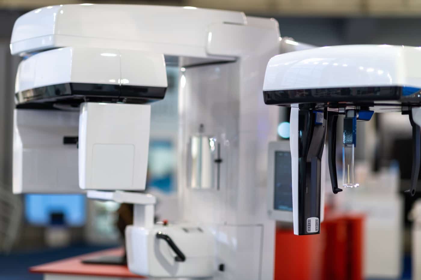
3D imaging: seeing the whole picture
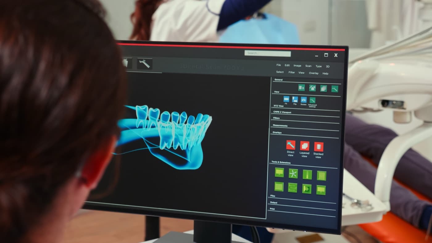
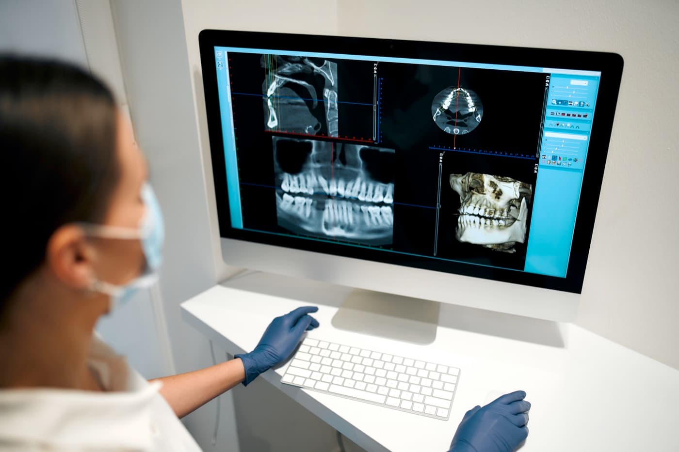
A standard dental X-ray is a flat photograph. Cone-beam CT (CBCT) imaging is a three-dimensional model of your jaw — and for surgical planning, that difference matters.
Why it matters
What 3D imaging lets your surgeon see
Bone in three dimensions
Height, width, and density at an implant site — measured, not estimated from a flat image.
Nerve position
The nerve canal in the lower jaw, mapped before wisdom-tooth removal or implant placement near it.
Sinus anatomy
The floor of the sinus above upper back teeth — central to sinus-lift and upper implant planning.
Impacted teeth, precisely located
The exact position and orientation of an impacted canine or wisdom tooth, and its relationship to neighboring roots.
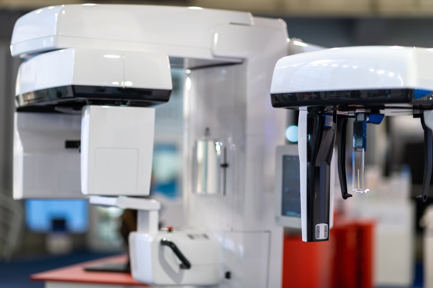
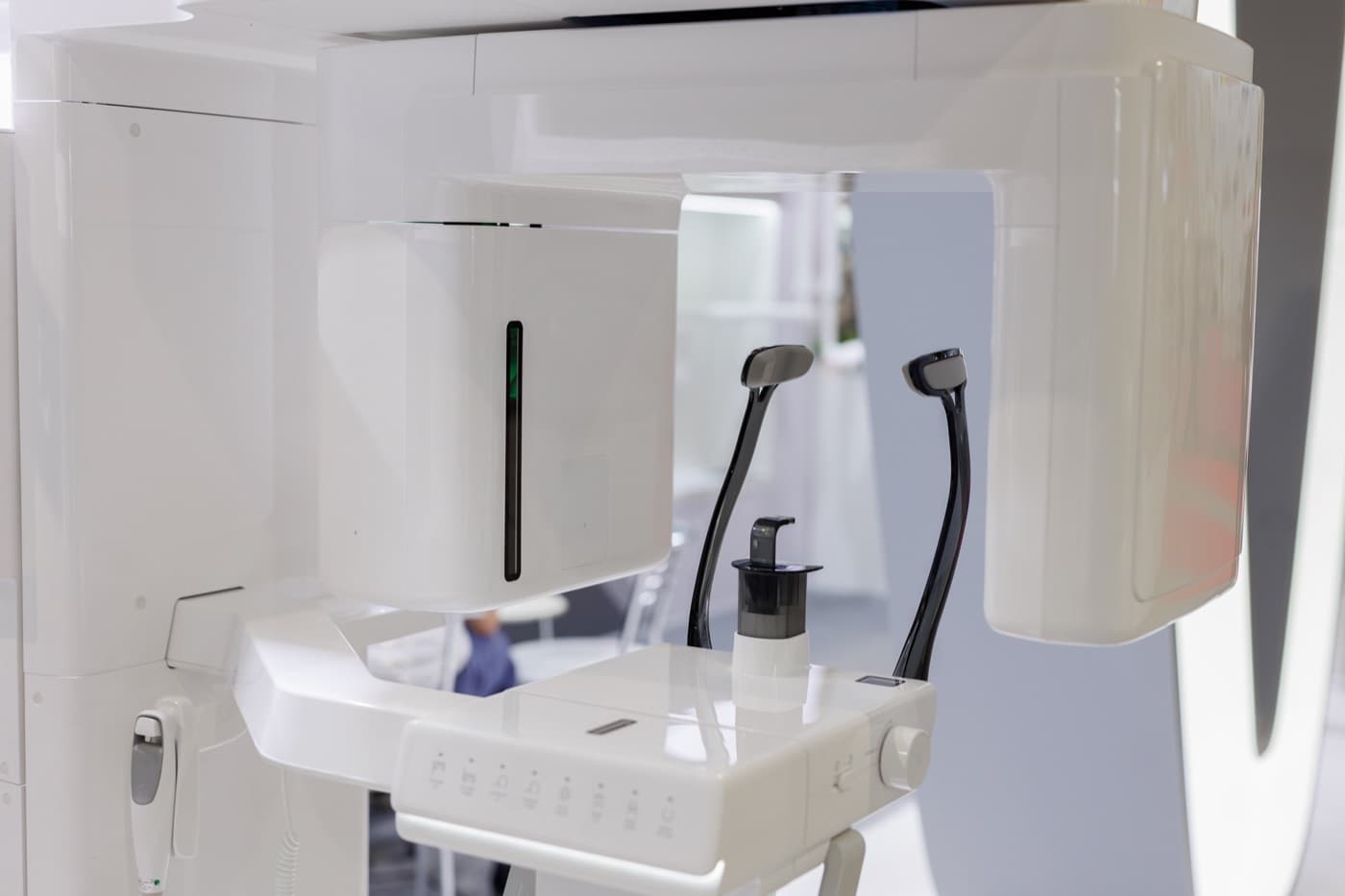
The scan itself is quick and non-invasive: you sit or stand while the imaging arm rotates around your head. Nothing touches you, and the images are available to review immediately — often at the same visit.
Used with judgment
Imaging when it helps, not by default
Like all X-ray-based imaging, CBCT involves radiation exposure, and the professional standard is simple: image when the information changes the plan. Your surgeon recommends 3D imaging when your case needs it — complex anatomy, implant planning, proximity to nerves or sinuses — rather than scanning every patient by routine.
If you have questions about imaging and radiation — especially for younger patients — ask at your consultation. You should expect a direct answer about why a scan is or is not recommended for you.
Measured, not estimated.
A standard dental X-ray is a flat photograph. Cone-beam CT imaging is a three-dimensional model of your jaw — and for surgical planning, that difference matters.
Your surgeon recommends 3D imaging when your case needs it — complex anatomy, implant planning, proximity to nerves or sinuses — rather than scanning every patient by routine.
Where 3D imaging shows up in your care
Further reading
Patient education written for this practice on the questions that come up before and after this procedure.
Dental Implants
How 3D Imaging Decides Where a Dental Implant Should Go
Modern implant placement is planned in three dimensions before surgery begins. Here is what the scan shows and why it changes the plan.
5 min read
Technology & Imaging
What 3D Imaging Shows That a Flat X-Ray Can't
Three-dimensional imaging reveals depth, angle, and nearby anatomy that flat films compress into a single view. Here's why that matters before surgery.
5 min read
Technology & Imaging
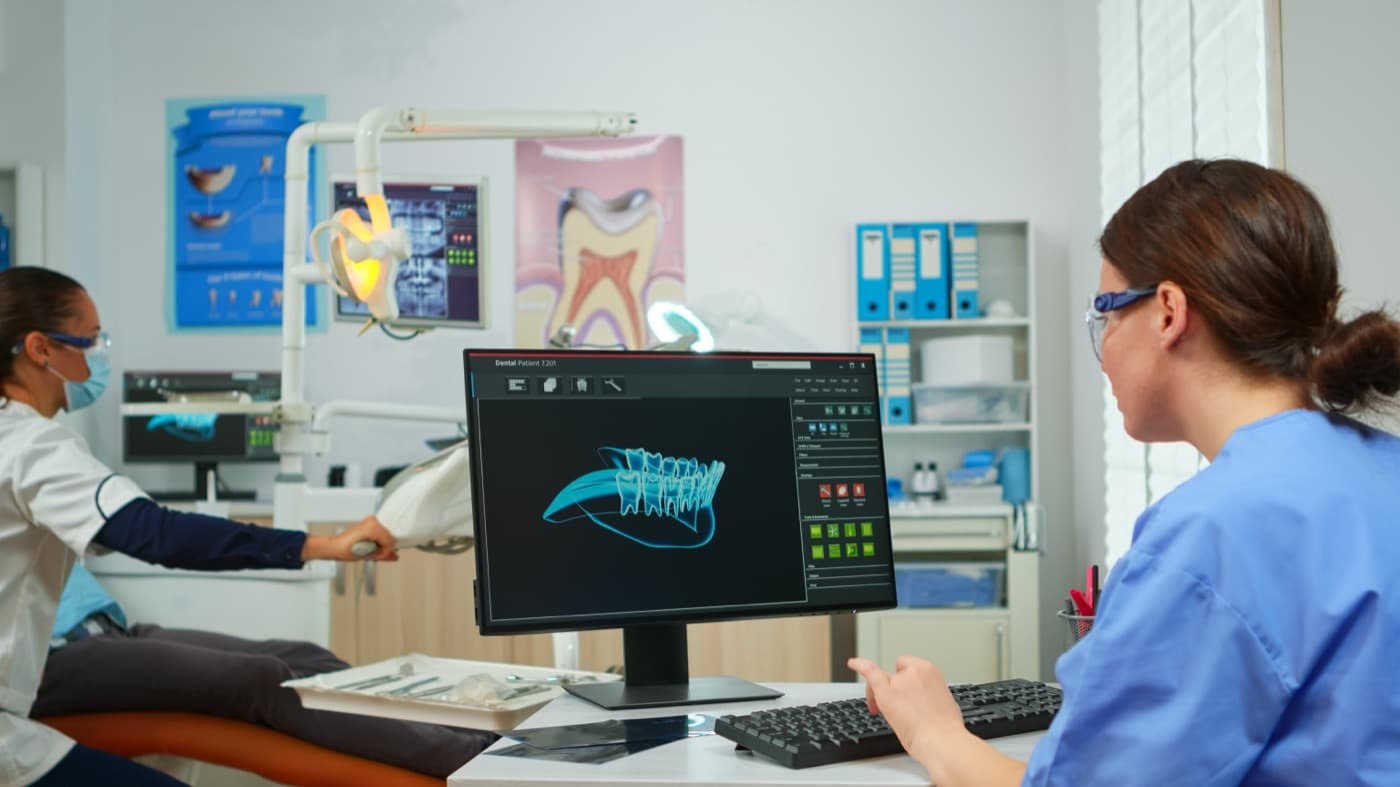
How Surgeons Plan Your Procedure Digitally — Before You're in the Chair
Much of modern oral surgery happens on a screen before it happens in an operatory. A look at how digital planning shapes the procedure you experience.
5 min read
Technology & Imaging
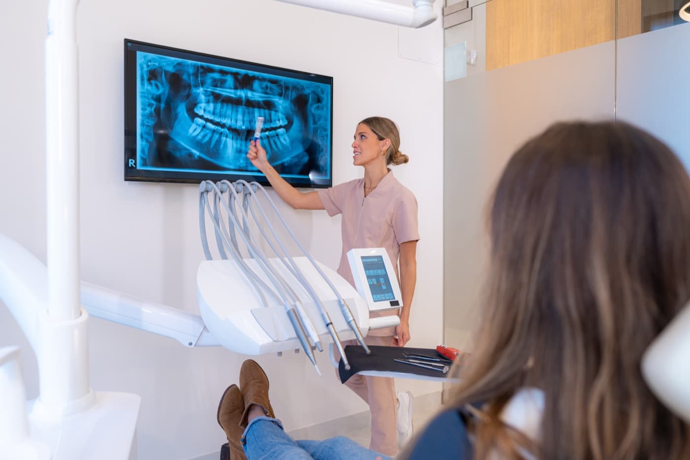
The Panoramic X-Ray: Why It's Often the First Image at a Surgical Consultation
One sweeping image of the whole jaw helps a surgeon see the full picture at a consultation. Here's what a panoramic X-ray shows and what it doesn't.
4 min read
Technology & Imaging
When Imaging Is — and Isn't — Recommended Before Oral Surgery
More imaging is not automatically better care. How oral surgeons decide when a scan is clinically indicated, and when an existing image is enough.
5 min read
Planning starts with seeing clearly
If your case calls for 3D imaging, it can usually be done here as part of your consultation.