
What 3D Imaging Shows That a Flat X-Ray Can't
5 minute read
DRAFT — pending clinical review v1.0-draft Clinical reviewer (to be assigned)
Three-dimensional imaging reveals depth, angle, and nearby anatomy that flat films compress into a single view. Here's why that matters before surgery.
A flat picture of a three-dimensional problem
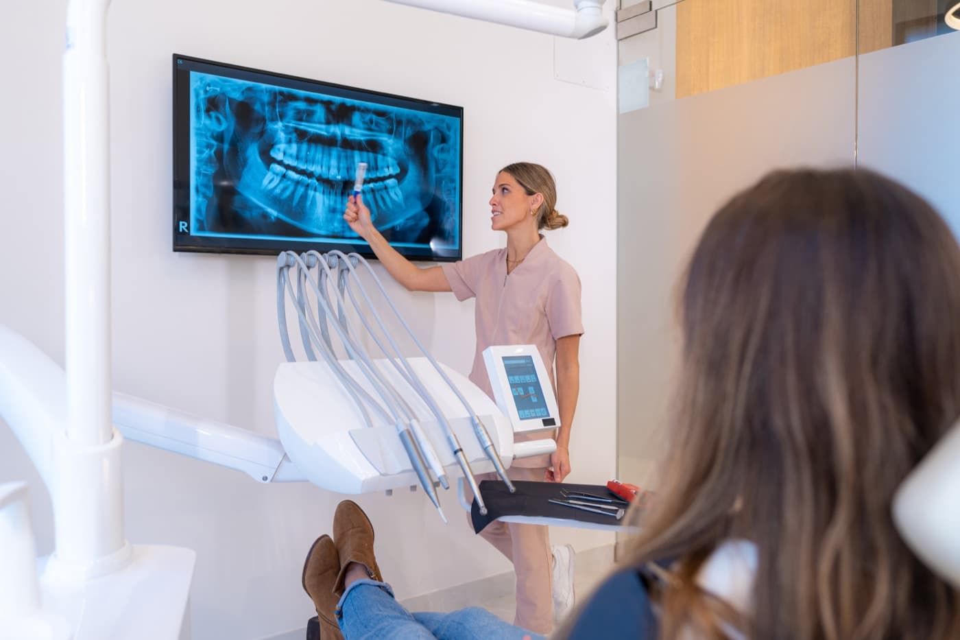
A traditional dental X-ray is a shadow. It takes everything in the path of the beam — teeth, bone, nerves, sinuses — and flattens it onto a single plane. That works well for many everyday dental questions, like spotting decay between teeth or checking the fit of a filling. But the mouth is not flat, and some of the most important surgical questions are about depth: how close is a tooth root to the nerve canal, or how thick is the bone behind a missing tooth?
On a flat film, two structures that sit at very different depths can appear to touch or overlap. A tooth root might look like it wraps around a nerve when it actually sits comfortably to one side. Three-dimensional imaging exists to resolve exactly this kind of ambiguity when the answer would change how a procedure is planned.
Seeing anatomy from any angle
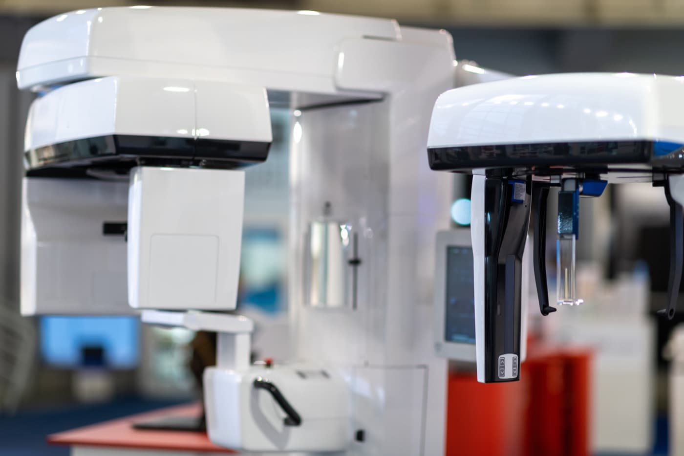
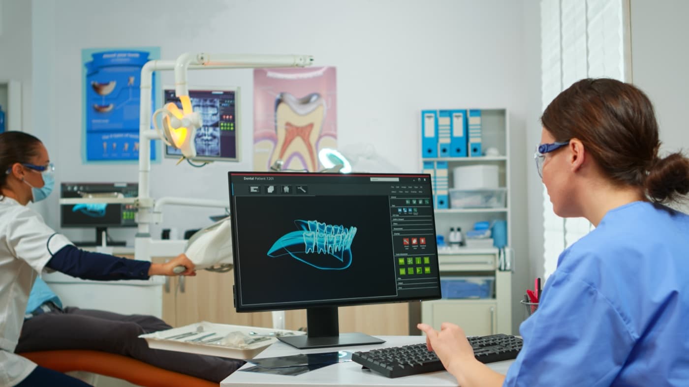
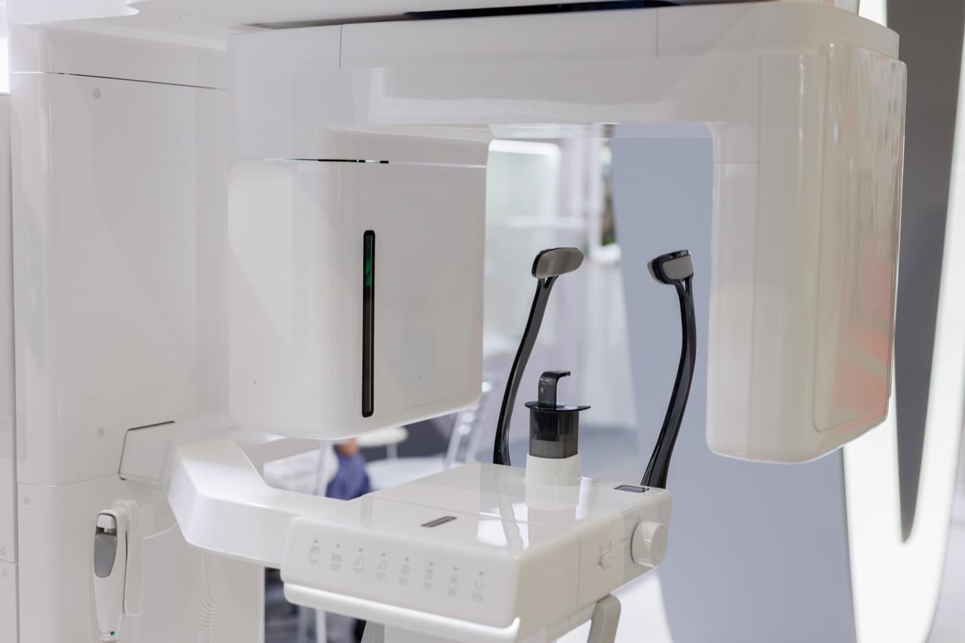
Cone beam imaging, the type of 3D scan most often used in oral surgery, produces a volume rather than a picture. A surgeon can slice through that volume in any direction — top to bottom, front to back, side to side — and rotate the view to trace a structure along its full path.
That freedom matters for questions a flat image simply cannot answer. Which side of the jaw does a nerve canal run on? Is an impacted tooth tipped toward the cheek or toward the tongue? Does the bone in a planned implant site have enough width, not just enough height? These are spatial questions, and a spatial image is the honest way to answer them.
Surgeons also use these views to plan the path of the procedure itself — where to make an opening, how to approach a buried tooth, and what to stay away from along the route. The image becomes less like a photograph and more like a map that can be explored from every direction.
Where the difference shows up in surgical care
Before removing a deeply positioned tooth, a surgeon may want to know its exact relationship to the nerve that supplies feeling to the lip and chin. Before placing a dental implant, the surgeon may want to see the true contour of the bone and the position of the sinus floor. Before treating a cyst or lesion, the surgeon may want to understand how far it extends and what it touches.
In each case, the value of the scan is not the technology itself — it is the specific clinical question it answers. When the question can be answered with a simpler image, a simpler image is the right choice.
3D imaging is a prescription, not a default
Not every patient needs a 3D scan, and not every procedure calls for one. Oral surgeons prescribe imaging when it is clinically indicated — when the added information is expected to confirm or change the plan for your care. If a standard X-ray answers the question, that is usually where imaging stops.
If your surgeon does recommend a 3D scan, it is reasonable to ask what question the scan is meant to answer. A good answer will be specific: the position of a nerve, the shape of the bone, the boundaries of a lesion. That conversation is part of informed care.
What to take away
Flat X-rays and 3D scans are complementary tools, not competitors. One gives a fast, familiar overview; the other resolves depth and detail when the stakes call for it. Your surgeon's job is to match the tool to the question.
This article is educational and is not a substitute for an examination. Whether imaging is appropriate for you — and which kind — is a decision made with your surgeon based on your own anatomy and treatment plan.
Common questions
Will I automatically get a 3D scan before oral surgery?
Can I use a scan taken at my dentist's office?
Does a 3D scan hurt or require preparation?
This article is general education, not personal medical advice. It cannot account for your health history or imaging, and it never replaces the evaluation and written instructions you receive from your surgeon.
Ready to talk through your plan?
Request a consultation and our team will follow up to schedule your visit.