
The Panoramic X-Ray: Why It's Often the First Image at a Surgical Consultation
4 minute read
DRAFT — pending clinical review v1.0-draft Clinical reviewer (to be assigned)
One sweeping image of the whole jaw helps a surgeon see the full picture at a consultation. Here's what a panoramic X-ray shows and what it doesn't.
One image, the whole story
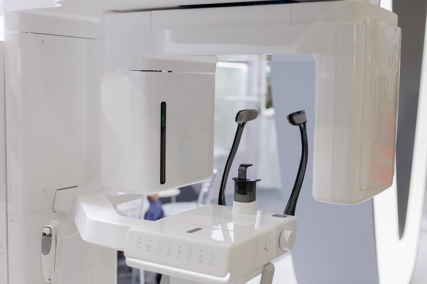
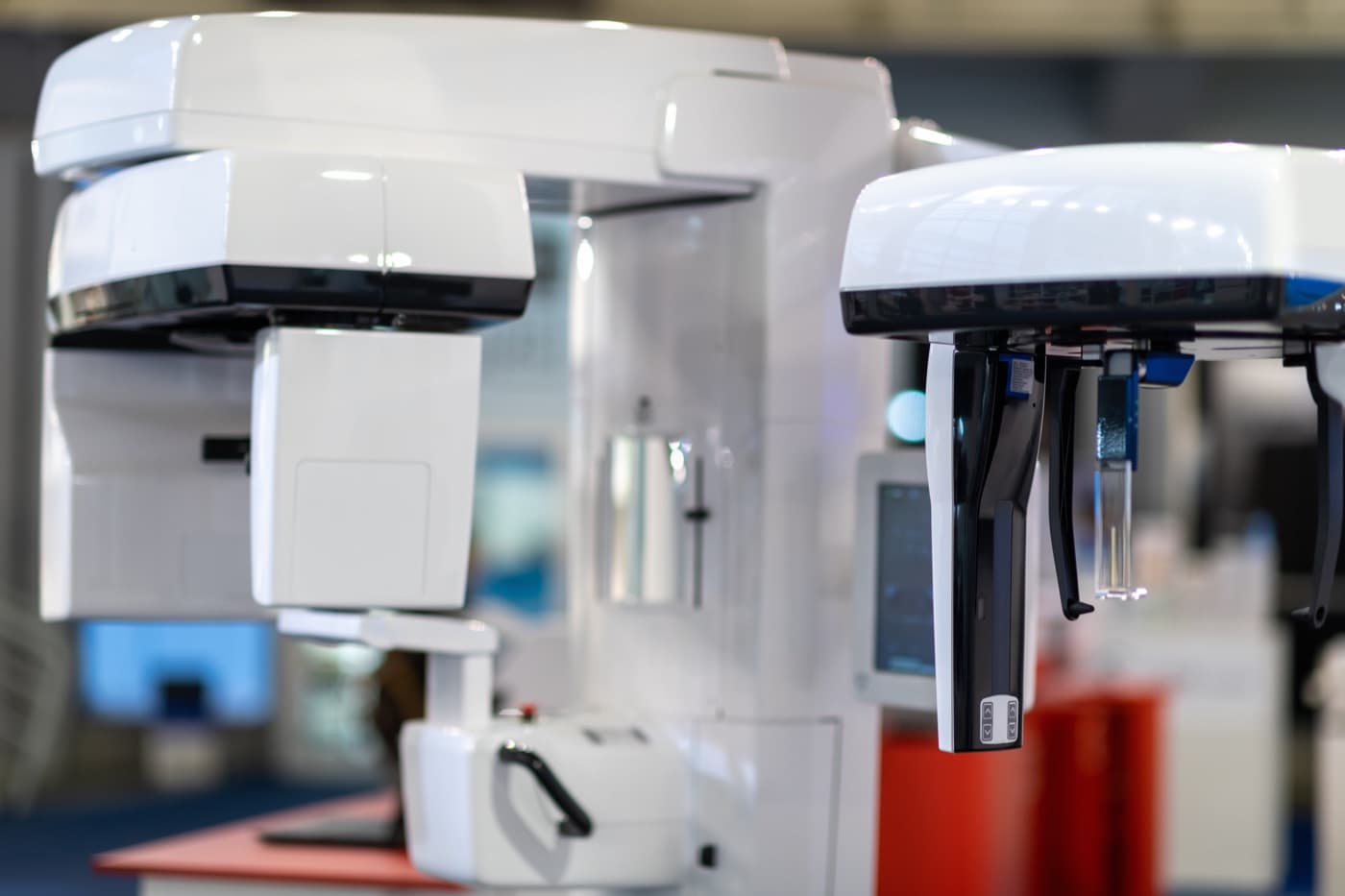
A panoramic X-ray captures the entire upper and lower jaw in a single sweep — every tooth, both jaw joints, the sinuses, and the surrounding bone, all on one image. The machine rotates around your head while you stand or sit still, and the result is a wide, curved view of the whole region an oral surgeon cares about.
That breadth is why the panoramic image is so often the starting point of a surgical consultation. Before focusing on the one tooth that brought you in, the surgeon can survey everything around it: the neighbors, the opposing teeth, the bone levels, and anything unexpected hiding elsewhere in the jaw.
The image is captured quickly and comfortably. Nothing is placed inside your mouth, which makes the panoramic approach easier for patients with a strong gag reflex, limited jaw opening, or significant pain — exactly the circumstances that often bring people to an oral surgeon in the first place.
What a surgeon reads in a panoramic image
For wisdom tooth evaluations, the panoramic view shows how the teeth are positioned, how developed their roots are, and how they relate — at least in broad strokes — to the nerve canal in the lower jaw and the sinuses in the upper jaw. For extractions and implant consultations, it gives an overall sense of bone quality and the health of surrounding teeth.
It can also reveal things no one was looking for: an impacted tooth that never erupted, a cyst forming around a buried crown, or changes in the bone that warrant a closer look. Part of the value of a wide image is precisely that it does not limit the surgeon's view to the tooth on the referral slip.
Because the image covers both jaws at once, it also helps the surgeon see how a planned procedure fits your mouth as a whole — how removing one tooth might affect its neighbors, or how a future implant would relate to the teeth it has to work alongside.
What it doesn't show — and when more is needed
A panoramic X-ray is still a flat, two-dimensional image, and it involves some built-in distortion because of the way it is captured. It shows relationships side to side and top to bottom, but not depth. If a wisdom tooth root appears to overlap the nerve canal, the panoramic image alone cannot say whether they actually touch.
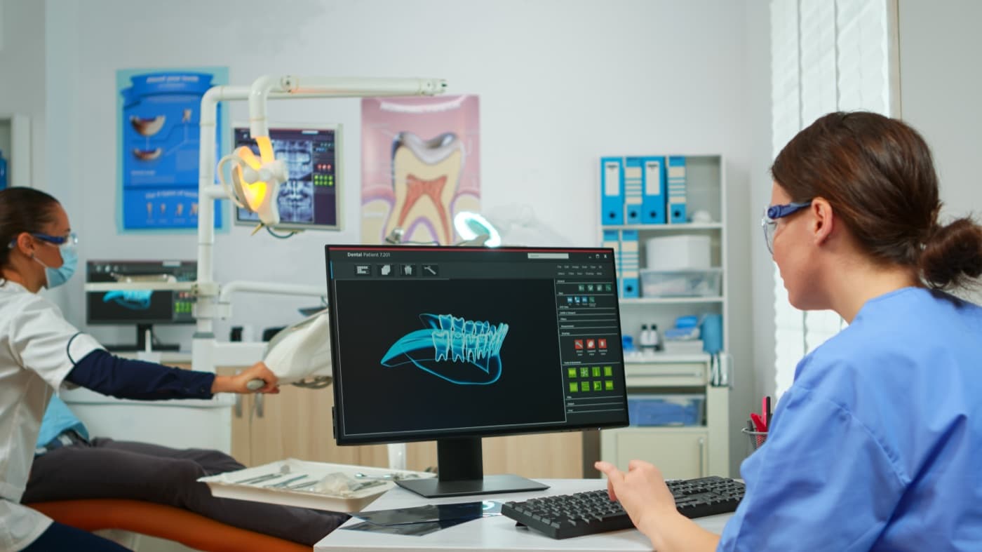
When that kind of question matters to the safety of a procedure, the surgeon may prescribe three-dimensional imaging as a next step. The panoramic image identifies the question; the 3D scan, when clinically indicated, answers it.
This is not a shortcoming so much as a division of labor. Each type of image has a role, and responsible imaging uses the simplest tool that answers the question at hand.
Practical notes for your visit
If your dentist has taken a recent panoramic image, ask to have it sent to the surgeon's office before your consultation — surgeons prefer to reuse recent imaging when it still answers the clinical question, and only prescribe new images when they are needed.
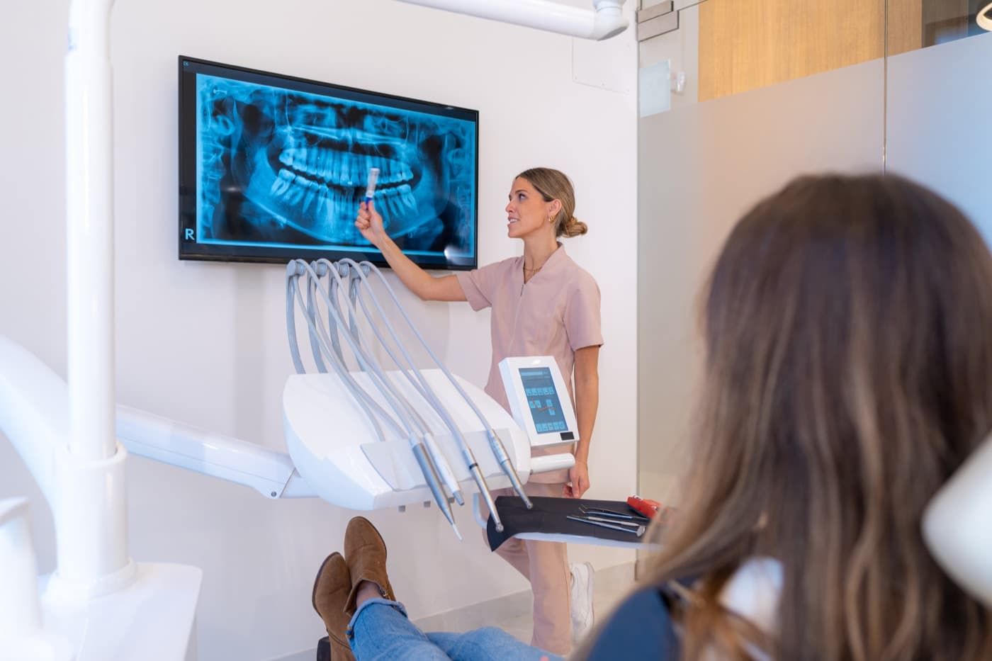
During the consultation, ask the surgeon to point out what they see. Most are glad to trace the anatomy with you on screen, and understanding your own image makes every later conversation about treatment easier.
If you feel uneasy about imaging or unsure why an image is being taken, say so. The team can explain the purpose of each step, and understanding what a picture is for usually makes the process feel routine. This article is educational and is not a substitute for a clinical examination.
Common questions
Why take a panoramic image if I might need a 3D scan anyway?
Do I need to do anything to prepare for a panoramic X-ray?
My referral came with an image from my dentist. Will it be reused?
This article is general education, not personal medical advice. It cannot account for your health history or imaging, and it never replaces the evaluation and written instructions you receive from your surgeon.
Ready to talk through your plan?
Request a consultation and our team will follow up to schedule your visit.