
How 3D Imaging Decides Where a Dental Implant Should Go
5 minute read
DRAFT — pending clinical review v1.0-draft Clinical reviewer (to be assigned)
Modern implant placement is planned in three dimensions before surgery begins. Here is what the scan shows and why it changes the plan.
Why a flat X-ray is not enough
A traditional dental X-ray shows your jaw from one angle, flattened into a single image. It can reveal a great deal, but it cannot show how wide your bone is, exactly where a nerve travels, or how close the sinus floor sits to a planned implant site. Those are three-dimensional questions.
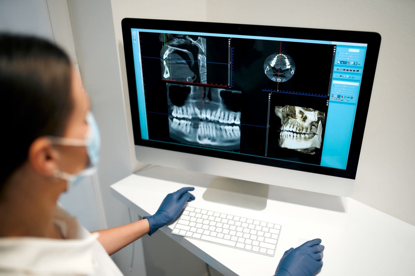
Cone-beam computed tomography — often shortened to CBCT — answers them. The scanner circles your head and produces a detailed volume that your surgeon can rotate, slice, and measure on screen from any direction.
If you have questions about imaging safety, ask them freely. The field of view is limited to the region your surgeon needs, and your team can explain exactly what your scan involves and why it is worthwhile for your plan.
What the surgeon looks for in the scan
First, the bone itself: its height, width, and density at the implant site. An implant needs to be surrounded by sound bone on all sides, and the scan shows whether that envelope exists or whether grafting should come first.
Second, the structures to respect. In the lower jaw, a nerve canal carries the nerve that gives feeling to your lip and chin. In the upper jaw, the sinus cavity sits above the back teeth. Planning in three dimensions lets your surgeon position implants with clear margins around these landmarks.
The scan also reveals things a surface exam cannot: the true angle of the bony ridge, hidden defects left by an old extraction or infection, and the character of the bone the implant will engage. Each detail nudges the plan toward a safer, more predictable placement.
Planning backward from the final tooth
The best implant position is not simply where bone is thickest — it is where the final tooth needs support. Digital planning software lets your team place a virtual tooth in its ideal position first, then work backward to the implant angle and depth that will support it.
This is where coordination with your restorative dentist matters. The scan can be merged with digital models of your teeth and bite so that surgical decisions serve the end result you will actually see and use.
Working backward this way protects appearance as well as function. An implant that emerges through the gum in the right spot lets the crown look like it grew there; one placed without that discipline can force the restoration into awkward compromises.
From the screen to the surgery
Once a plan is set, it can travel into the operating room in several ways. Some surgeons use a printed surgical guide — a custom template that fits over your teeth or gums and directs the implant to the planned position and angle. Others use the plan as a detailed map alongside their clinical judgment.
Whichever method is used in your case, the principle is the same: the important decisions were made deliberately, in advance, with full information — not improvised at the chair.
Digital planning also improves the conversation with you. Instead of describing anatomy in the abstract, your surgeon can turn the screen around and show you your own jaw, the planned implant, and the reasoning — which makes your consent genuinely informed.
What this means for you as a patient
Three-dimensional planning is a reason implant recommendations sometimes change after the scan. A site that looked fine on a flat film may show thin bone in cross-section, or the reverse. If your plan shifts, ask to see the images — most surgeons are glad to show you exactly why.
The scan itself is brief and does not touch you. Your surgeon will determine whether and when imaging is needed as part of your individual evaluation. Bring your questions about the technology to the visit; seeing your own anatomy on screen tends to answer most of them better than any article can.
Common questions
Is a CBCT scan uncomfortable?
Do all implant patients need a 3D scan?
What is a surgical guide?
This article is general education, not personal medical advice. It cannot account for your health history or imaging, and it never replaces the evaluation and written instructions you receive from your surgeon.


Ready to talk through your plan?
Request a consultation and our team will follow up to schedule your visit.